


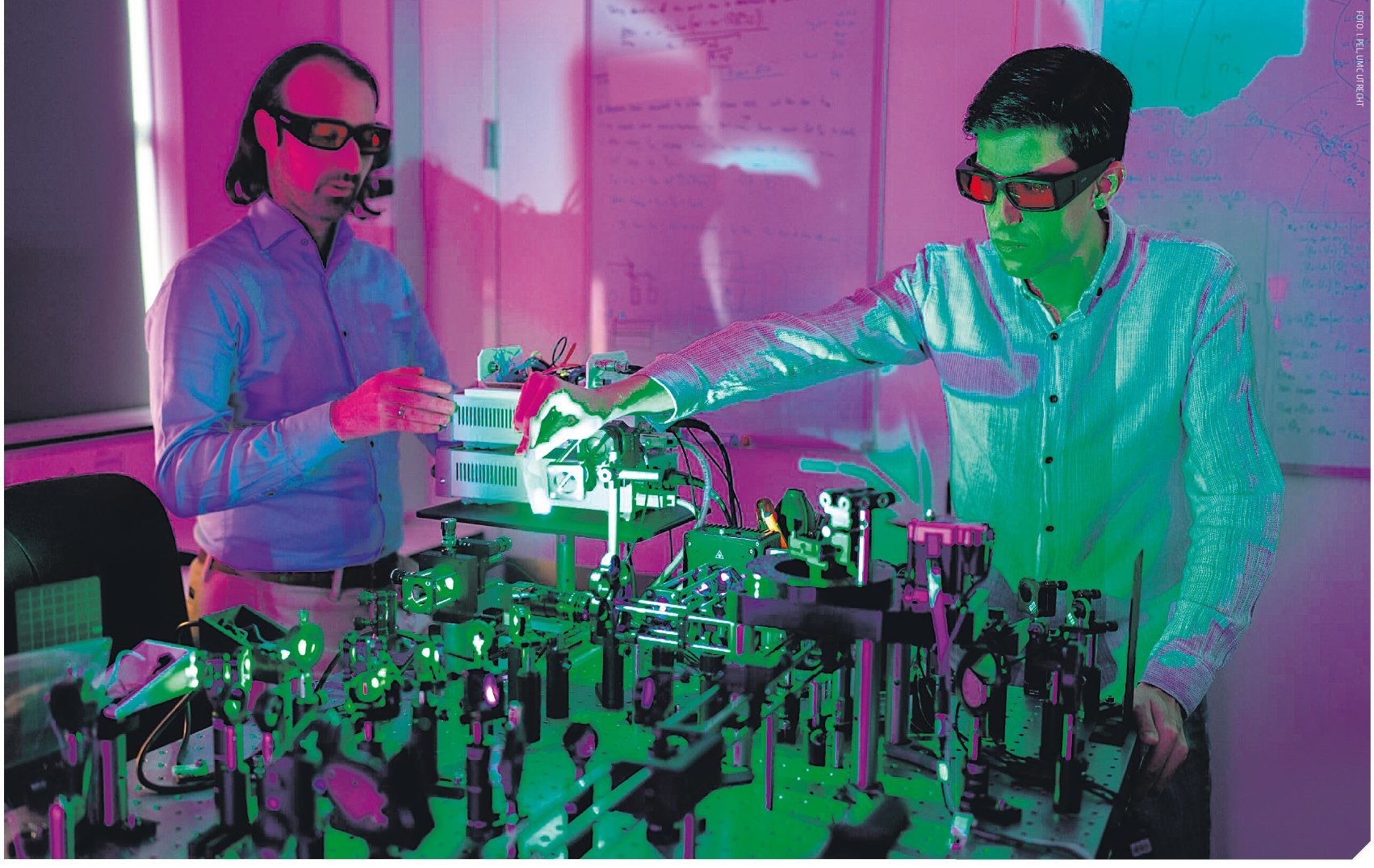
 Riccardo Levato and Sammy Florczak (a PhD student in his team) working on the volumetric printer. Photo: I. Pel, UMC Utrecht
A tiny liver or a gleaming sliver of pancreas rolling straight off the printer. The breakneck development of bioprinters promises a future in which life-saving tissues for transplantation can be built at the touch of a button.
The image seems futuristic: gleaming machines printing living tissues and fragments of organs in a matter of minutes. Yet this has not been mere fantasy for some time now. In laboratories around the world, researchers are developing 3D printers that extrude soft materials laden with living cells. Beating pieces of cardiac muscle, smooth chunks of cartilage, mini-livers or sections of pancreas – all of them emerging from what are known as bioprinters. The aim is eventually to transplant these printed structures into the body, where they could support failing organs or take over other bodily functions. In the Netherlands, too, this work is currently in full swing.
‘Within Europe, the Netherlands is a pioneer when it comes to bioprinting’, says biomedical scientist Jos Malda of UMC Utrecht and Utrecht University. He works on printing tissue for the knee joint. With his group, he is developing osteochondral implants, which they intend to transplant not only in humans but also in horses. Their laboratory houses several 3D printers, each contributing to a different component. ‘First we create an anchor for the bone from a kind of elastic ceramic’, Malda explains. ‘We then print a collagen-like hydrogel containing cells on top of that. Finally, we combine the whole thing with ultra-thin fibres that act as a kind of Kevlar, enabling the cells to withstand the forces in the knee joint.’
Although the knee implants are not yet ready for use in patients, Malda and his group are currently working hard towards testing them in horses. Osteochondrosis is a condition in young horses in which a small area of bone beneath the cartilage in the knee has developed abnormally. ‘There are no good treatments for it yet’, says Malda. ‘The dream is to be able to deploy our implant as a last resort. What we learn from that will probably also be applicable to humans, and could therefore represent a first step towards a clinical application.’
Hydrogel
The common thread in bioprinting is that printing is done with what is known as a bioink. This typically consists of a hydrogel containing cells and substances that the body will accept. The trick is to cure this hydrogel into the desired structure, with cells fixed in precise positions. Biomedical engineer Riccardo Levato, a colleague of Malda who is likewise affiliated with UMC Utrecht and Utrecht University, has optimised a technique for this purpose – so-called volumetric 3D printing. Using beams of light, he is able to grow mini-organs in a specific shape within the gel (see box: volumetric 3D printing). Levato: ‘Thanks to this technique, we can print various structures, such as mini-livers and pieces of pancreas. We use these to study how such tissues respond to different drugs. That helps to reduce animal experiments and costly clinical trials, since we can already rule out the majority of non-working drugs in the laboratory.’
Eindhoven University of Technology also presented a new technique in January – Xolography (capitalised because it is a brand name) – capable of printing complex biological structures at high resolution (Advanced Materials, 2025) (see box: Xolography). Biomedical engineer Miguel Dias Castilho, the group leader behind the research, uses the technique to print so-called scaffolds. ‘These are structures in which cells can settle and divide’, Dias Castilho explains. ‘After transplantation into the body, the scaffolds could serve as a literal scaffold to help tissues grow.’
It might sound as though these breakthroughs place us only a few steps away from printing fully functional organs, but Malda knows better. ‘Printing cells in the shape of an organ has been possible for some time, but that does not give it functionality. You can print a heart, but there is no switch to make it beat.’ It is precisely this functionality that represents the great challenge for the future of bioprinting, as Levato also sees it: ‘For a working tissue, cells not only need to be in the right position – they also need to be able to communicate with one another. This cell communication only develops after printing. To make progress, we therefore need to focus not only on the technology, but also on the biology.’
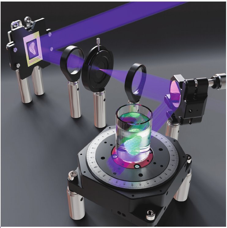 A simplified 3D representation of the volumetric printer. Illustration: S. Florczak, Levato Lab, UMC Utrecht and Utrecht University
To improve communication between cells, Levato is currently working on refining the materials used in printing. ‘The molecules present in the gel are highly influential in determining how cells respond’, says Levato. ‘In our current research, we are trying to develop a gradient of molecules within the gel in order to guide cells in this way.’ Dias Castilho is also studying how cell function can be influenced. He does this by designing different scaffolds with a variety of shapes. ‘We are investigating how mechanical forces and the geometry of the scaffold can direct stem cells to differentiate into tissue-specific cells’, he explains. ‘Here we focus in particular on bone and muscle tissue, because these cells are highly sensitive to mechanical forces.’
Burns
The fact that fully functional organs cannot yet roll off the printer does not mean that bioprinters have failed to achieve practical results. In France, the first human clinical trials for the transplantation of bioprinted skin in patients with severe burns are currently under way. In addition, an American patient received a transplant of a printed ear in 2022, made from their own cartilage cells. ‘Although such an ear, despite its high functionality, has a relatively low complexity’, says Malda. ‘It contains no blood vessels, for instance. This simplicity is why cartilage is currently regarded as a promising material for printing structures.’
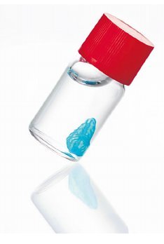
An image of a piece of pancreatic tissue, printed with the volumetric printer. Photo: E. Cohas
Levato observes that the focus in the research field is currently not on printing entire organs, but on parts of them. Levato: ‘The larger the organ, the exponentially more complex it will be to print. I foresee that in the future we will more likely print pieces of tissue that take over a single function of an organ, such as regulating glucose in the pancreas by assisting with insulin production.’ Dias Castilho also doubts whether we should expect beating hearts or functioning kidneys from the printer any time soon. ‘Although I do think there is something wonderful about having such a moonshot goal’, he says. ‘Beyond that, I believe our current scaffolds can already go a long way towards improving therapies. If our work were to lead to new strategies for treating patients with muscular diseases – because we better understand how cells respond to their environment – that would already be more than enough for me.’
Volumetric 3D printing
Although volumetric 3D printing was originally conceived for plastics and non-biological materials, Riccardo Levato and his colleagues further developed the technique in 2019 to print living cells. They use the technique to grow mini-organs in a test tube containing gelatine. This gel, derived from collagen found in the body, solidifies when exposed to specific light beams from a laser. To fix cells in the desired position, it is therefore important to illuminate the gel in precisely the right places. The researchers achieve this by rotating the test tube during exposure. ‘We project hundreds of different 2D scans of the mini-organ from different angles onto the gel to create a 3D object’, explains Levato. The cells obstruct the light in this process. The researchers solve this using a substance called iodixanol, which temporarily makes the cells transparent without harming them. By washing away the gel that has ultimately not reacted, only the printed structure remains. To print complex mini-organs consisting of multiple tissue types, the researchers combine their technique with extrusion – the ‘traditional’ 3D printing technique used, among other things, to print plastic objects by building them up layer by layer. ‘We start, for example, with extrusion to print a ring of cells, during which we can rapidly and efficiently swap between different tubes of cells. We then use volumetric 3D printing to print blood vessels from a different cell type around and through this structure, and to give the tissue its final shape.’
Xolography
Miguel Dias Castilho uses Xolography to build scaffolds for cells. This technique was devised by two German researchers who founded their own spin-off company, Xolo. When Xolo began looking, four years ago, for a way to apply their technique to printing complex biological structures, Dias Castilho decided to collaborate with them. Like volumetric 3D printing, Xolography projects 2D light patterns of the desired print structure onto the gel to cure it. The main difference between the two techniques is that Xolography uses not one but two light sources: UV light and visible light. The UV light ‘activates’ the gel, after which it becomes receptive to projections of visible light that cure it. The desired structure is printed only where the two light sources converge within the gel. This allows the technique to print large volumes at high resolution in just a few minutes. With a resolution of 20 micrometres, Dias Castilho hopes to build constructs with the right geometry to attract endogenous cells once implanted in the body. The idea is that, following transplantation, cells will be able to settle into the scaffold and divide there. As they grow, the cells would gradually break down the scaffold, so that ultimately only healthy tissue remains. ‘To achieve this goal, we are first looking in the lab for the mechanical signals needed to accelerate this process in vivo.’